Disease Overview
Disc degeneration reduces the height of the disc and may cause a Herniated disc. The vertebrae of the backbone are cushioned by intervertebral discs that act as shock-absorbers and allow frictionless movement of your back. It is made up of a soft gel-like center called the nucleus pulposus that is surrounded by a tough outer ring of annulus fibrosus. A herniated disc is a condition in which the nucleus pulposus bulges out through the damaged or broken annulus fibrosus. Herniated disc is also called bulging disc, ruptured disc or slipped disc. Disc herniation causes compression of the spinal cord and/or spinal nerves. Spinal cord compression can cause pain in the arm and legs. In rare cases, it can lead to permanent damage and even paralysis.
What is Artificial Cervical Disc Replacement?
Artificial cervical disc replacement is a spine surgery to replace a degenerated (deteriorated) disc in the neck with an artificial disc. The artificial disc, like the natural healthy disc, is used to replace the degenerated disc. It restores the height between the two cervical vertebrae, enlarging the neural foramen (nerve passageway in the spine) and relieving the pressure on the spinal nerves. This stabilizes the cervical spine and restores normal mobility of the neck.
How is Artificial Cervical Disc Replacement Performed?
For the procedure, the cervical spine is approached through an incision in the front of the neck. The affected disc is identified with the help of imaging studies and removed. The artificial disc is then placed precisely in the disc space between the vertebrae. After checking the range of motion of the neck and confirming the proper fit of the artificial disc, the incision is sutured closed.
Risks and Complications of Artificial Cervical Disc Replacement
As with any surgery, artificial cervical disc replacement may be associated with certain complications such as infection, bleeding and nerve injury causing temporary hoarseness of the voice and difficulty in swallowing.
Prodisc L
Anterior Lumbar Total Disc Replacement
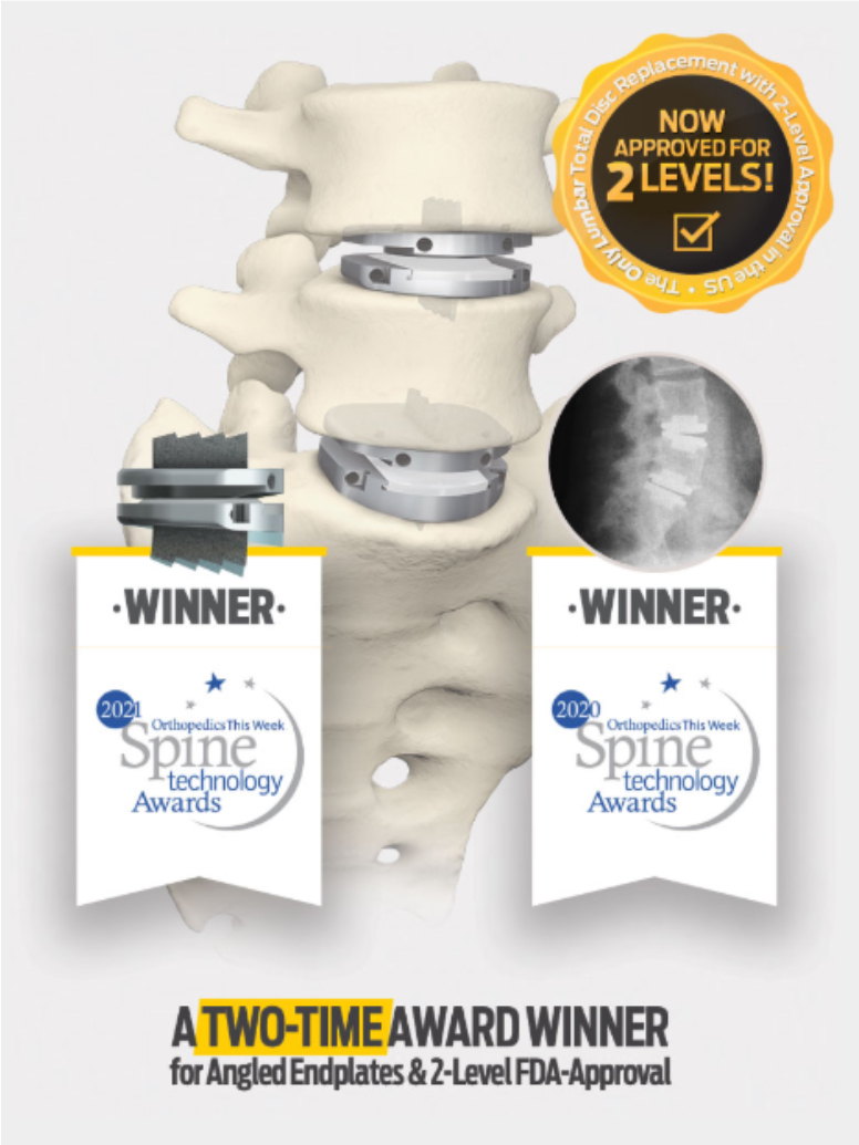
The most studied and clinically proven total disc replacement (TDR) technology in the world is now the only total disc replacement system in the U.S. approved for two-level use in the lumbar spine.
Anatomic Endplates™
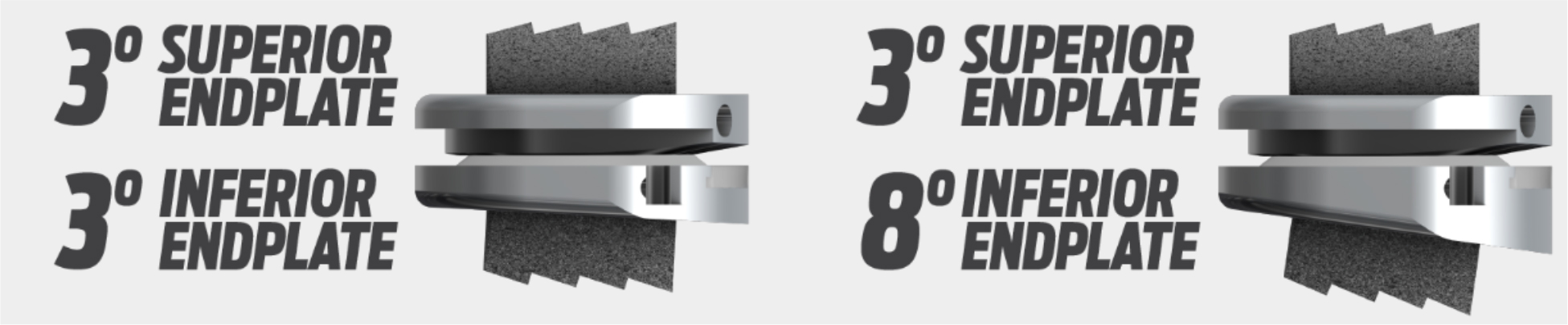
The prodisc L Total Disc Replacement system now has a greater selection of endplates available. These unique endplates have been designed to shift the lordotic angle of the implant to the inferior endplate, expanding the options available to surgeons to better address the varied lumbar anatomy and pathology of patients.
The Most Studied TDR System in the World
Beginning with clinical usage in 1990, the prodisc design has been validated with over 225,000 device implantations worldwide and more than 540 published papers. 1
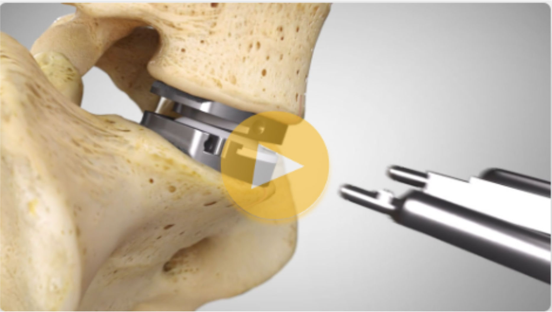
See the prodisc L Surgical Technique
Determined Safe & Effective for Degenerative Disc Disease
The prodisc L Total Disc Replacement has been proven safe and effective for treating degenerative disc disease (DDD) at two levels from L3 to S1.
The prodisc L Surgery is intended to:
- Remove the diseased disc
- Restore normal disc height
- Reduce discogenic pain
- Potentially provide motion in the affected vertebral segment
Design Philosophy
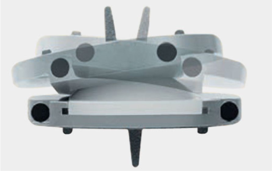
The prodisc L implant has been designed to maintain the physiological range of motion in the spine. The implant was developed using the clinically proven ball and socket concept used in joint replacement implants for over 40 years. The prodisc L implant is composed of three components – two cobalt chrome alloy (CoCrMo) endplates and an ultra-high molecular weight polyethylene (UHMWPE) inlay.
Mechanism of Action
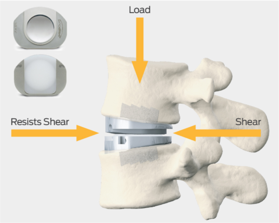
The prodisc implant is a ball and socket design with a fixed center of rotation. This patented design has been in clinical use since 1990 and utilized across the entire product platform. The fixed center of rotation allows physiological range of motion while providing stability to the spine and significantly reducing reoperations at the adjacent levels.
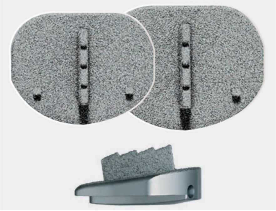
Secure Fixation
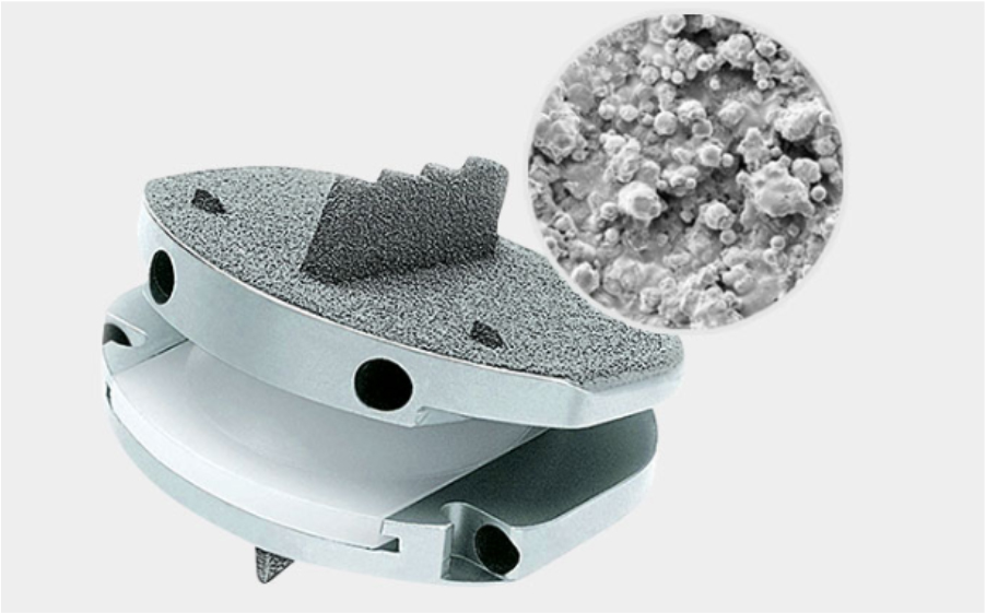
- Patented central keel and lateral spikes provide secure primary fixation
- Plasma-sprayed titanium surface on bone contacting surfaces promotes integration
Anatomical Sizing
The prodisc L system is available in 12 anatomical combinations, including:
- Medium and large footprints
- Heights: 10mm, 12mm, 14mm
- Lordotic angles: 6° and 11°
Safe & Reproducible Surgical Technique
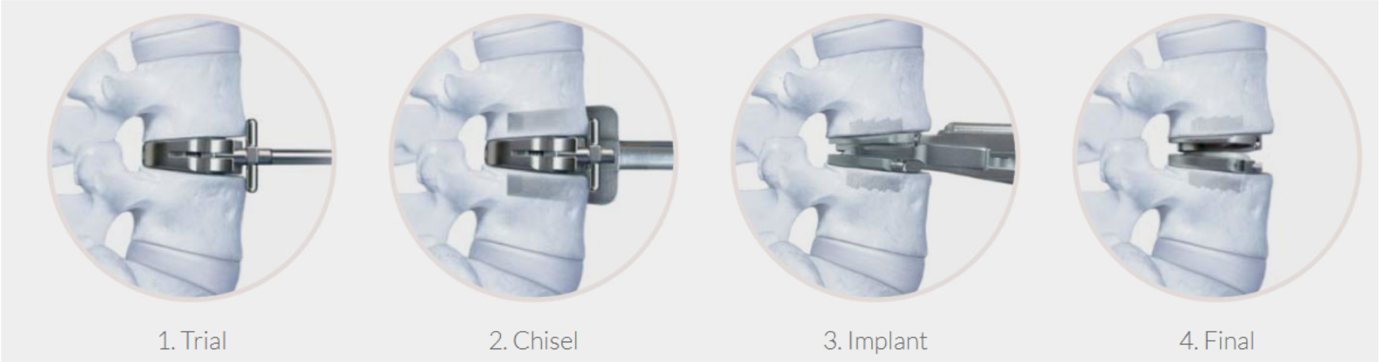
Working with leading spine surgeons from around the world, the prodisc L instrumentation and surgical technique has been refined to facilitate safe and reproducible implantation through a midline, mini-open anterior approach to the lumbar spine.
- Three-Step Implantation Process
- Enables accurate sizing and precise placement of the implant
Streamlined Instrumentation
- Designed for a midline, mini-open anterior approach
- Clear visualization into the disc space
- Minimizes exposure & reduces risk of vascular damage
Reference: Search performed on PubMed, Embase, and Ovid Medline® (1988–2020).